Photoacoustic imaging is a non-destructive medical imaging method developed in recent years. It combines the high contrast characteristics of pure optical imaging with the high penetration depth of pure ultrasound imaging to provide high resolution and high contrast tissue imaging.
The small animal photoacoustic imaging system developed by Endra of the United States has nano-molar sensitivity and high resolution of 280um, which can detect photoacoustic signals below 20mm. It can also be used for quantitative analysis of molecular imaging of small animals. At the same time, it combines near-infrared detection technology to enhance the absorption signal of light, which in turn increases the contrast of the picture, making the experimental results clearer and more accurate.
Microvascular system imaging and quantitative analysis
Near-infrared light has a deep penetration ability in biological tissues; and the injection of the exogenous dye indocyanine green as a light absorption enhancer increases the selective absorption of light by blood vessels, thereby enhancing vascular light. The intensity of the acoustic signal. Indocyanine green (ICG), also known as indocyanine green, is an FDA-approved light-absorbing contrast-enhanced infrared sensitizing dye. ICG is rapidly infused with hemoglobin after intravenous injection into the body, and is rapidly distributed along the blood circulation. Systemic intravascular. Because of its high binding rate to hemoglobin and its ability to be absorbed by extrahepatic tissues, ICG is a highly effective vascular marker dye that has been widely used in angiographic studies and tumor detection.
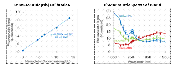
Since the concentration of ICG and its light absorption value are linearly related, the hemoglobin concentration marked by it can also be obtained by conversion of the light absorption value. The left graph of Figure 1 shows the proportional relationship between the hemoglobin concentration and the light absorption value of the same sample. The image to the right shows the determination of oxygen saturation of different samples under a series of near-infrared spectroscopy excitations. At the same time, ICG can also be used as a contrast enhancer to increase the contrast of the image and assist in imaging.
Recently, researchers have also used nano-gold particles with strong emission signals in the near-infrared spectrum as contrast agents to image the cerebral cortical vasculature, which can clearly reveal 100um microvessels.
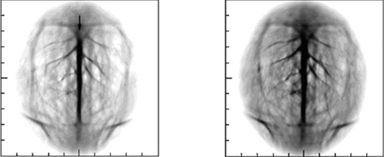
Figure 2 shows the brain vascular imaging before (left) and after injection (right) injection of nano-contrast agent. It can be seen that the cortical microvasculature can be clearly obtained after injection .
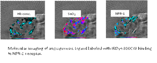
In addition, Endra can also do quantitative analysis by means of labeling. The figure below shows the calculation of blood oxygen content in blood vessels and the quantification of dye-labeled receptor content.
Fizzy Bath Bombs,Bath Bomb,Healcier Oem Bath-Bomb,Nail Buffer Hota
Dongguan Vanilla Bioengineering Co., Ltd. , https://www.healthecigarette.de