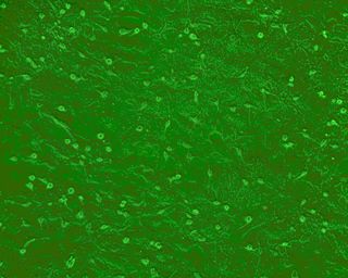
Normal mouse primary brain astrocyte culture

PriCells - normal mouse primary brain astrocyte culture
First, experimental reagents
1. Medium: PriCells Medium + 10% FBS + 1% P/S + PriCells Supplement
2. Cryopreservation solution: PriCells Medium + 20% FBS + 10% DMSO
3. Washing solution: 1 × PBS (pH 7.4 ) + 1% P/S
4. Staining solution: 0.4% Trypan Blue
5. Digestive juice: PriCells Isolation of Primary Cell Kit
6. Detection reagent: anti-mouse CD 11b/c antibody, fluorescently labeled secondary antibody, ethanol-acetone mixture (1:1)
Second, the experimental equipment
1. Petri dish
2, culture bottle
3, direct cut and eye scissors
4, ophthalmology and hemostats
5, 10ml syringe
6, glass dropper
7, beaker
8, 15ml centrifuge tube
Third, the experimental process
Material
↓
Remove the brain's meninges and blood vessels, remove the hippocampus
↓
Rinse 3 times in 1 × PBS (pH 7.4) to remove the superficial bloodshot
↓
First use the ophthalmology to tear the tissue into a braid, cut it with an eye scissors for 10 minutes, and then gently pipette 20 to 30 times with a pipette.
↓
Transfer the tissue to a 15ml centrifuge tube and add the digestive juice (PriCells)
↓
Place the tube to 37 ° C, 15-20 min water bath constant temperature digestion
↓
Stop the reaction by adding digestion stop solution (PriCells)
↓
Centrifugation, 1000rmp, 10min, discard the supernatant
↓
Resuspend, count cells
↓
Inoculation density is 1 × 10 6 /ml
↓
Culture at 37 ° C, 5% CO 2
Fourth, experimental operation
1. The culture bottle is pre-coated. The flask was pre-packaged one day before the test, placed in an ultra-static table or dried in an oven at 45 ° C (remember to keep it sterile), placed in a refrigerator at 4 ° C, and set aside.
2. Material: normal Kunming mice (within 24 hours after birth).
3. Material pretreatment: After the mice are sacrificed, they are immersed in a volume fraction of 75% ethanol for disinfection, and the vascular clamp is used to clamp the neck from behind. The scalp and the skull are separated from the occipital portion by the ophthalmic scissors, and then the ophthalmology is followed. Peel off and remove meninges and blood vessels. The brain (excluding the hippocampus) was placed in 1 × PBS (pH 7.4), rinsed to the surface of the blood, and the brain tissue was milky white.
4, digestive juice: use the ophthalmology to tear the tissue into a braid, then use the ophthalmic scissors to cut repeatedly for 10min, and finally use a pipette to gently blow 20 to 30 times, the shredded tissue is transferred to a 15ml centrifuge tube with a gun In the 37 ° C water bath digestion for 15 min.
5, stop digestion: add digestion stop solution according to 1:1, stop the enzyme reaction.
6. Collect the resuspended cells: centrifuge at 1000 rpm for 10 min, discard the supernatant, resuspend the culture medium, and stain the cells.
7. Culture cell density: The cell density was adjusted and inoculated into the culture flask at 1 × 10 6 /ml.
8. Culture: Place in a 37 ° C, 5% CO 2 incubator.
V. Cell identification
1. Microscopic identification: Under the phase contrast microscope, the cells are star-shaped, and the cell density is increased, and the cobblestone-like mosaic arrangement is arranged, which has the characteristics of contact inhibition. The cells have good light transmission.
2. Immunohistochemical identification: Immunofluorescence staining was performed using OX-42 (CD 11b/c).
3. Cell climbing. The washed coverslips were placed in a 6-well culture plate, and the cells were seeded at 3×10 4 cells/well. At 48 hours, the cells were overgrown, and the coverslips covered with cells were taken out with tweezers for use.
4. Cell fixation: The cells were washed with PBS, then placed in a mixture of ethanol and acetone, fixed for 10 min, and naturally dried in the air.
5. Specific antibody: Dilute the antibody according to the dilution requirements, add the antibody, and incubate at 37 ° C for 60 min.
6. Wash: Wash 1 × PBS (pH 7.4) 3 times × 15 minutes, and dry.
7. Labeled antibody: Add fluorescently labeled antibody and incubate at 37 ° C for 30 min.
Washing: Wash 1 × PBS (pH 7.4) 3 times × 15 minutes, and dry.
8. Cover: Seal the tablet.
9. Microscopic examination: Under the fluorescence microscope, the glial cell material showed strong yellow-green fluorescence, especially around the nucleus.
Six, matters needing attention
1. Select materials to select newborn Kunming mice (within 24 hours). The process of taking the material should be as sterile as possible, and the meninges, blood vessels and hippocampus should be removed as much as possible.
2, pay attention to eliminate residual blood on the surface of brain tissue as much as possible, to avoid the influence of blood cells and certain serum components on the adhesion of glial cells.
3, glial cell separation damage seriously affects the growth of glial cells, so the enzyme digestion concentration and digestion time should be strictly controlled.
4. Strictly control the culture conditions of glial cells. Including the quality and concentration of cell culture fluids (culture medium, growth factors, serum, antibiotics).
5. Regarding the culture of the bottle or not, there is literature research showing that there is no particularly large difference between the coated and uncoated. In the experiment, it is possible to consider whether or not to coat.
6. The inoculation amount of the subculture cells was 1 × 10 6 (25 cm 2 flask), and the cell doubling time was 36 hours. Cell subculture is best for 5-8 generations, the number of passages is increased, the cell volume is increased, the gap between cells is increased, the cells are firmly attached, and the passage time is prolonged.
Seven, PriCells cell picture

1. We have Good location

We`re located in the well known Ningbo International Port.

2. We`re Well-experienced

Tropical Group has many factories in the world, has 48 years experience in seafood export

3. We`re High quality assurance

Our company has passed HACCP, BRC, MSC, FDA, KOSHER and the other certifications.

4. We can do Best price

We are an industry and trade integration enterprise, which producing, packing and selling all by ourselves, greatly reducing the price difference caused by broker

5. We have Rich products

We have our own design department and R & D team, which can design personalized products according to customers' requirements.

6. We can do On-time delivery

Our products are exported to the United States, South America, Africa, Europe, Russia and other countries and regions.  Company exports amounted to more than US$60 million in last year, enjoys high reputation in the aquatic products industry.

Contact: Ms. Sunny Wang

Canned Sardine

Canned Sardine,Sardine Canned,Sardine Fish Canned,Canned Sardine With Vegetables

Tropical Food Manufacturing (Ningbo) Co., Ltd. , https://www.tropical-food.com